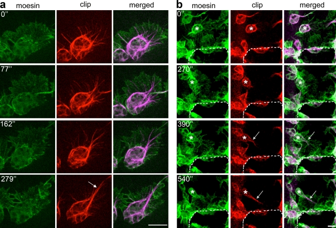
Figure 2.
Microtubules are transiently bundled within the lamellae of migrating hemocytes. (a) Live imaging of a hemocyte expressing GFP-Moesin (actin) and mCherry-CLIP170 (microtubules) revealed dynamic microtubules rapidly bundling into an arm (arrow) to polarize the cell’s morphology. (b) After laser ablation, a hemocyte (asterisks) in the vicinity of the wound extends a microtubule arm (arrows) before acquisition of a polarized lamellar morphology. The dashed lines indicate the wound edge. Time is shown in seconds. Bars, 10 µm.

