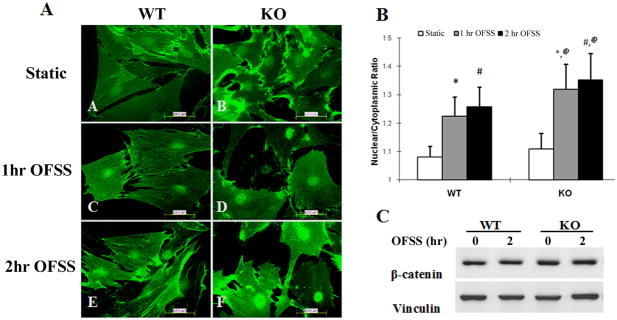
Figure 1.
Nuclear translocation of β-catenin in response to OFSS is enhanced in Nmp4 KO primary calvarial osteoblasts compared to osteoblasts isolated from WT mice. (A) Immunofluorescence microscopy of primary calvarial osteoblasts subjected to static culture conditions or OFSS for 1 hour or 2 hour (12 dynes/cm2). Slides were fixed immediatelyand processed for immunofluorescence using an antibody against β-catenin followed by FITC-conjugated secondary antibody. Results shown are using cells isolated from N6 mice. Hoechst 33342 was used to label and identify the outline of nuclei (not shown). (B) Quantification of the nuclear/cytoplasmic ratio of β-catenin in cells isolated from N6 mice. A minimum of 100 cells in each treatment condition were selected randomly and the fluorescent grey value was calculated in the nucleus and in an identically sized area near the nucleus in the cytoplasm. Hoechst labeling was used to define the nuclear outline. The 2-way ANOVA indicated a strong genotype effect (p<0.0001), a strong treatment effect (p<0.0001) and a genotype × treatment interaction (p<0.0001). The Tukey’s HSD post hoc analysis determined that WT STATIC=KO STATIC<WT 1HR FLOW=WT 2HR FLOW<KO 1HR FLOW=KO 2HR FLOW. *p<0.0001 1h OFSS vs. static; #p<0.0001 2h OFSS vs. static; @ p<0.0001 1 hr OFSS KO vs. 1hr OFSS WT and 2 hr OFSS KO vs. 2 hr OFSS WT. NOTE: all treatment groups of the F2-KO cells exhibited a significantly greater nuclear:cytoplasmic ratio of β-catenin than that observed in the F2-WT cells (not shown). (C) Immunoblot analysis for total cellular β-catenin (normalized to total cellular vinculin) from cells maintained in static culture or subjected to OFSS for 1 hr (not shown) or 2 hr. No change in total β-catenin levels were detected as a consequence of OFSS.

