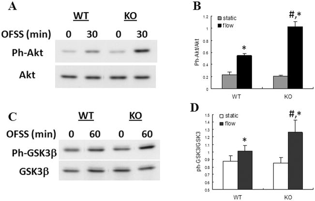
Figure 3.
Activation of AKT and GSK3β in response to 30 and 60 min of OFSS, respectively, was enhanced in Nmp4 KO osteoblasts compared to WT controls. (A) Representative immunoblot of total cellular protein from Nmp4 and WT osteoblasts maintained in static culture or subjected to OFSS for 30 min and probed either with antibody specific for phosphorylated Akt or for total Akt. (B) Quantification of immunoblots illustrated in (A), n=3. The static vs. 30 min OFSS experiment was repeated three times with comparable results. *p<0.05 vs. static; #p<0.05 vs. WT flow. (C) Representative immunoblot of total cellular protein from Nmp4 and WT osteoblasts maintained in static culture or subjected to OFSS for 60 min and probed either with antibody specific for phosphorylated GSK3β or for total GSK3β. (D) Quantification of immunoblots illustrated in (C), n=3. The static vs. 60 min OFSS experiment was repeated three times with comparable results. *p<0.05 vs. static; #p<0.05 vs. WT flow.

