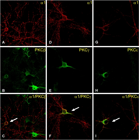
Fig. 6.
PKC isoforms are colocalized with GABAA receptor α1 subunits in ethanol-naive cells. Cortical neurons were stained with anti-α1 and PKCβ, PKCγ, or PKCε antibodies for confocal microscopy. A, D, and G, α1-labeled cells; B, E, and H, PKCβ-, -γ-, and -ε-labeled cells, respectively. C, F, and I are merged images of dual immunostaining of α1 subunit and the PKC isoform. The yellow color in merged panels (arrow) indicates colocalization of the two proteins.

