Fig. 2.
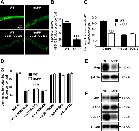
P-glycoprotein expression and transport activity are reduced at the blood-brain barrier of hAPP mice. A, representative images of brain capillaries isolated from 12-week-old wild-type and hAPP mice. Capillaries were incubated with 2 μM NBD-CSA, a fluorescent P-glycoprotein-specific substrate, for 1 h alone or with PSC833. B, specific (PSC833-sensitive) luminal NBD-CSA fluorescence after image analysis of brain capillaries. C, luminal fluorescein-hAβ42 fluorescence in brain capillaries from wild-type and hAPP mice. D, luminal fluorescence of the MRP-specific, fluorescent substrate, sulforhodamine 101, in brain capillaries alone (control) or with mannitol (osmotic tight junction disruptor), LTC4 (MRP inhibitor), NaCN (metabolic inhibitor), PSC833 (P-glycoprotein inhibitor), RAP (LRP1 inhibitor), or FTC (BCRP inhibitor). Data in B–D are mean ± S.E.M. for 10 capillaries from one preparation (pooled tissue from 10–20 mice per group). Shown are arbitrary fluorescence units (scale 0–255). ***, significantly lower than control, P < 0.001. E and F, Western blots for P-glycoprotein (P-gp) (E) and LRP1, RAGE, and GLUT-1 (F) of brain capillary membranes from wild-type and hAPP mice. β-Actin was used as protein loading control (pooled tissue from 20 mice per group).

