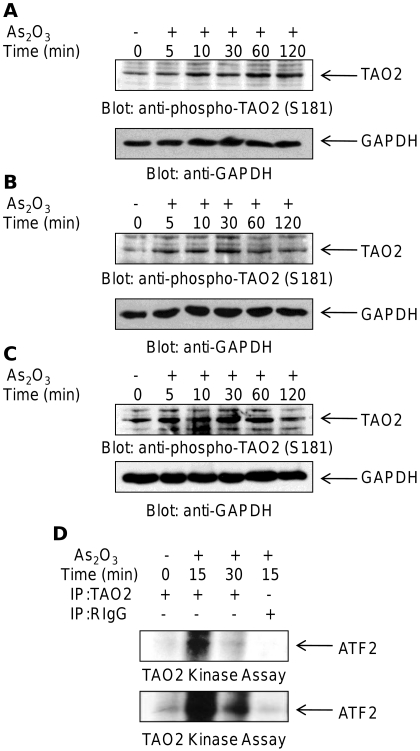
Fig. 1.
As2O3-dependent phosphorylation of TAO2 in leukemic cell lines. A, U937 cells were incubated in the absence or presence of As2O3 (2 μM) for the indicated times. Equal amounts of total cell lysates were resolved by SDS-PAGE and immunoblotted with an anti-phospho-TAO2 (Ser181) antibody (top). The same blot was reprobed with an anti-GAPDH antibody to control for protein loading (bottom). B, as in A, but using NB4 cells. C, as in A, but using NB4.306 cells. D, U937 cells were incubated with As2O3 (2 μM) as indicated. Cell lysates were subjected to in vitro kinase assays using activating transcription factor 2 as an exogenous substrate. Proteins were resolved by SDS-PAGE, and phosphorylated proteins were detected by autoradiography (top). Longer exposure of the same membrane is also shown (bottom).

