Fig. 8.
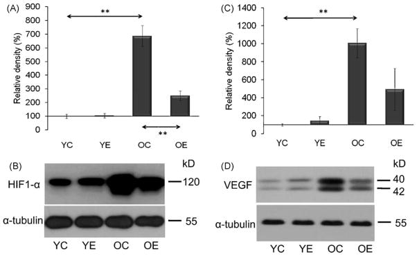
Aging increased the level of HIF-1α (A and B) and VEGF (C and D) and these increases were significantly attenuated by exercise training. Densities of the bands were normalized to tubulin which served as an internal control. In panels (A)–(D): YC, young control; YE, young exercised; OC, old control; OE, old exercised. Values are means ± S.E. for six animals per group; **p < 0.01.

