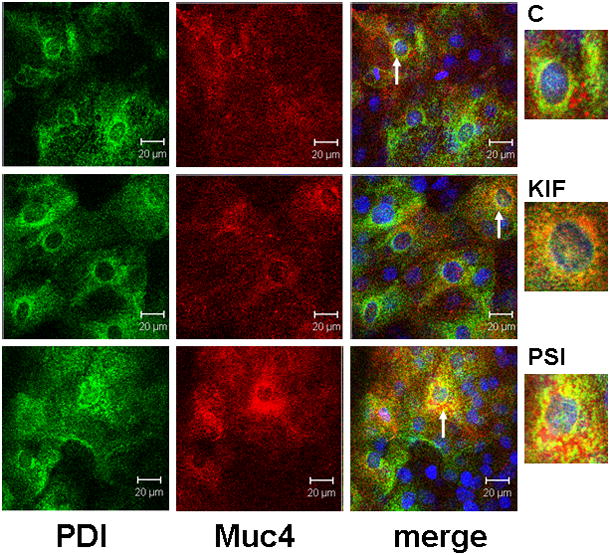
FIGURE 7.

ER localization of Muc4 in cells with blocked proteosomal degradation using XY- projection of three-color immunofluorescence staining. Cells were permeabilized according to the instructions with the SelectFX Alexa Fluor 488 endoplasmic reticulum-labeling kit. Proteins were double-labeled with polyclonal anti-Muc4 and with monoclonal anti protein disulphide isomerase (PDI). Muc4 was visualized with Alexa Fluor 594 (red) and PDI with Alexa Fluor 488 (green) antibodies. Nuclei were stained with DAPI. C – untreated cells; KIF – cells treated for 18 hours with 5 μM KIF, the α-mannosidase inhibitor; and PSI – cells treated for 4 hours with 0.01 μM PSI, the proteasome inhibitor. Insets show areas noted by arrows.
