
Fig. 1.
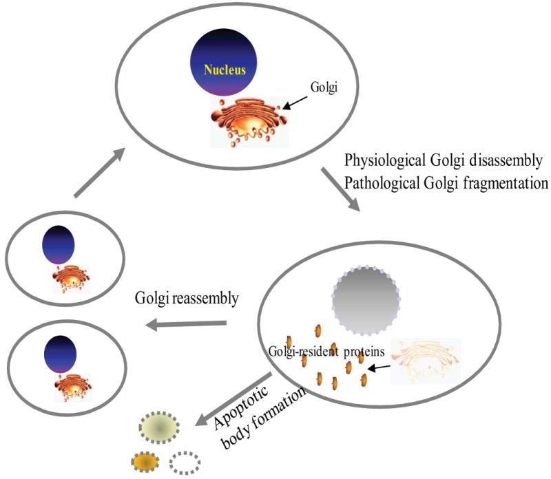
Schematic illustration of Golgi apparatus fragmentation and reassembly during cell mitosis and apoptosis. Many reasons can lead to Golgi fragmentation, but it can be generally classified as physiological Golgi disassembly (Mitosis) or pathological Golgi fragmentation due to apoptosis, presence of misfolded proteins, neurodegenerative diseases, Chlamydia infection, etc.