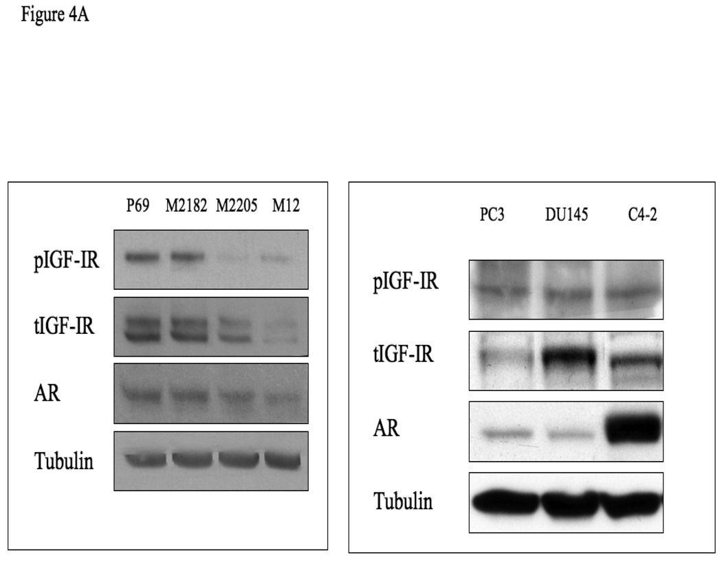
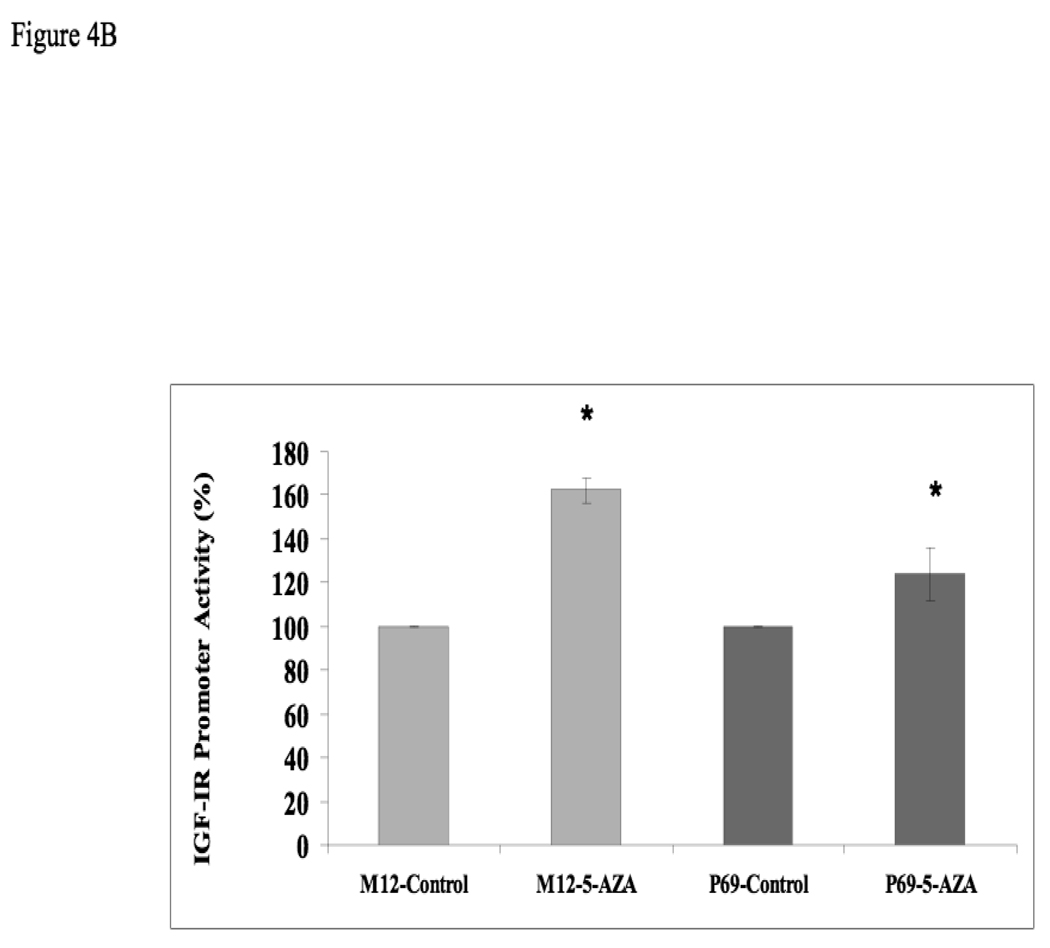
Figure 4. Expression of AR and IGF1R in prostate cancer cell lines.
(A) Cells were lysed in the presence of protease inhibitors, as indicated under Materials and methods. Equal amounts of protein (80 µg) were separated by 10% SDS-PAGE, transferred onto nitrocellulose filters, and blotted with anti-IGF1R, anti-phospho (p)-IGF1R, anti-AR, and anti-tubulin. (B) Effect of 5-Aza treatment on IGF1R promoter activity. P69 and M12 cells were treated with 5-Aza (or left untreated) and, after 24 h, cells were transiently transfected with a proximal IGF1R promoter-luciferase reporter construct [p(−476/+640)LUC], along with a β-galactosidase plasmid. Cells were harvested 48 h after transfection for luciferase and β-galactosidase assays. * p<0.02 versus respective control.


