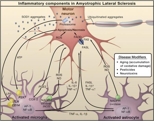
Figure 3. Inflammation in Amyotrophic Lateral Sclerosis.
The pathology of amyotrophic lateral sclerosis (ALS) is characterized by degeneration of motor neurons. Familial ALS is caused by mutations in the SOD1 gene, but the genes mutated in sporadic ALS are not yet defined. Progressive neurodegeneration of motor neurons in ALS may result from a combination of intrinsic motor neuron vulnerability to aggregates of mutant SOD1 protein and non-cell-autonomous toxicity exerted by neighboring cells. Toxic aggregates can induce inflammatory responses by microglia via Toll-like receptor 2 (TLR2) and CD14. Microglia can induce astrocyte activation by producing cytokines. Activated microglia and astrocytes amplify the initial damage to the motor neurons by activating AP-1 and NF-κB through production of proinflammatory cytokines and apoptosis-triggering molecules such as TNF-α and FASL. TNF-α and IL-1β exert neurotoxic effects in vitro, but deletion of the individual genes does not affect the course of the disease in an animal model. Dying motor neurons release ATP that can further activate microglia through the purinergic receptor P2X7 expressed by microglia.

