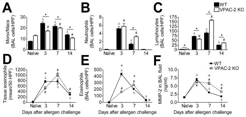
Figure 1.
Leukocyte infiltration was increased as a result of allergen challenge in both WT and KO groups. Monocytes/macrophages (A), neutrophils (B), lymphocytes (C), and eosinophils (E) in cytospun bronchoalveolar lavage (BAL) contents were differentially stained and counted. Peribronchovascular eosinophils were counted in 50 random HPFs in H&E stained sections (D). Total MMP-2 in the BAL was quantified via ELISA (F). All data are represented as the mean ± SEM, n=5 mice per group. Δ denotes p<0.05 of allergen exposed WT animals compared to their naïve controls, while # denotes p<0.05 of allergen exposed KO animals compared to their naïve controls. * indicates p<0.05 between WT and KO.

