Figure 4.
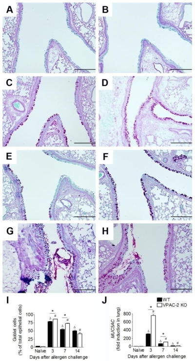
Representative photomicrographs of PAS-stained whole lung sections of naïve and allergen exposed WT (left panel) and VPAC2 KO (right panel) mice. Goblet cells (GCs) were scarce in naïve animals of WT (A) and KO (B) groups. GCs and mucus were evident in the airways of both groups at days 3 (C & D), 7 (E & F), and 14 (G & H). GCs were counted and reported as the percentage of total epithelial cells that lined the airways (I). The induction of MUC5AC mRNA in lung tissue was measured via real-time qPCR and reported standardized to naïve controls using the 2−ΔΔCt method. While both groups had MUC5AC induction in the allergic state, a marked difference between WT and KO levels occurred at day 3 (J). All data are represented as the mean ± SEM, n=3–5 mice per group. Δ denotes p<0.05 of allergen exposed WT animals compared to their naïve controls, while # denotes p<0.05 of allergen exposed KO animals compared to their naïve controls. * indicates p<0.05 between WT and KO. Scale bars = 200 μm

