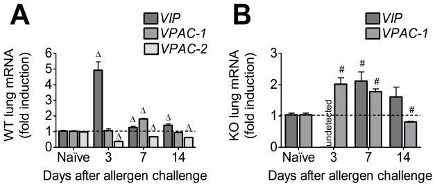
Figure 5.
Vasoactive intestinal peptide (VIP) and VPAC receptor mRNA in whole lung of WT (A) and VPAC2 KO (B) standardized to naïve controls using the 2−ΔΔCt method. VIP was markedly upregulated in WT at day 3 (A) while it was undetected in KO (B). Both groups had a similar expression pattern for VPAC1 (A & B), and the WT had a marked downregualtion of VPAC2 at all time points analyzed (A). All data are represented as the mean ± SEM, n=3 mice per group. Δ denotes p<0.05 of allergen exposed WT animals compared to their naïve controls, while # denotes p<0.05 of allergen exposed KO animals compared to their naïve controls.

