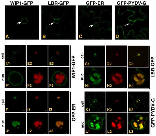
Fig. 8.
Single plane confocal micrographs of protein fusions of cells in which TagRFP-PYDV-M was co-expressed with various membrane markers fused to GFP by agroinfiltration in leaf epidermal cells of transgenic N. benthamiana plants. White arrows indicate the accumulation of markers on nuclear membranes in the absence of TagRFP-PYDV-M; A. WIP1-GFP (outer nuclear membrane marker), B. LBR-GFP (inner nuclear membrane marker), C. GFP-ER (endoplasmic reticulum) and D. GFP-PYDV-G (endomembranes). All remaining panels show coexpression of the membrane markers and TagRFP-PYDV-M. E1-3/F1-3, WIP1-GFP. G1-3/H1-3, LBR-GFP. I1-3/J1-3, GFP-ER. K1-3/L1-3, GFP-PYDV-G.

