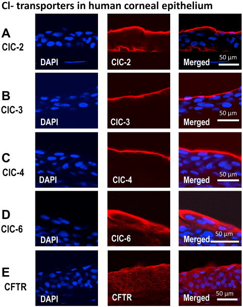
Figure 3. Confocal images of immunofluorescence staining for CLC family members in human corneal epithelium.
CLC family members CLC-2, CLC-3, CLC-4, CLC-6 and CFTR show distinct distribution in human corneal epithelium. Actin (not show here) and DAPI (for nuclear staining) were used as controls. (A), CLC-2 is distributed at the apical and basal layers of corneal epithelium. (B) and (C), CLC-3, and CLC-4 are expressed at the apical layer. (D) and (E), CLC-6 and CFTR expressed preferentially at the apical layers with expression level gradually decreasing towards the basal layer. Tear side is orientated to the top and basal side towards the bottom.

