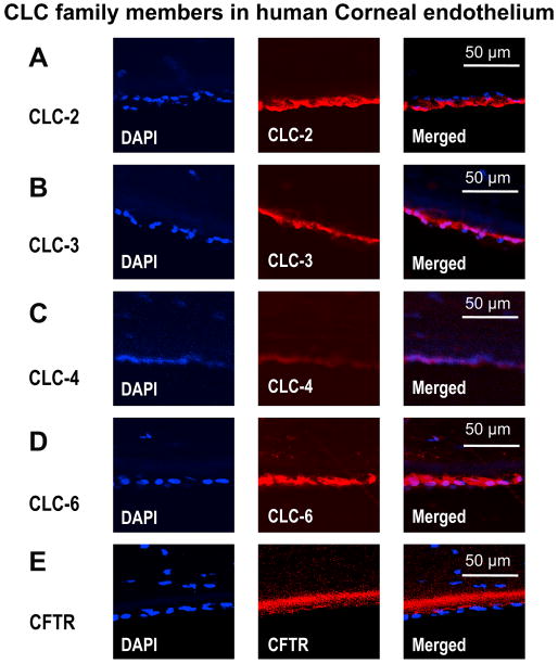
Figure 5. Confocal images of immunofluorescence staining for CLC family members in human corneal endothelium.
Human corneal endothelial cells are positive for immunostaining of CLC family members CLC-2, CLC-3, CLC-4, CLC-6 and CFTR. DAPI (for nuclear) staining was used to show the endothelium. Control staining using isoform IgG, or omission of the primary antibodies showed no staining (not show here). Stromal side is orientated to the top and apical side is orientated towards the bottom.

