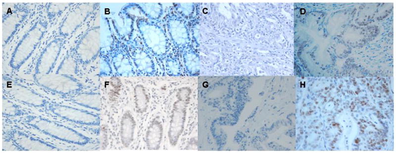
Fig. 2.

Immunohistochemistry for MLH1 and MSH2 protein expression (hematoxylin, 400x). (a) Normal colorectal tissue with no antibody staining for MLH1 (negative control). (b) Normal colorectal tissue with MLH1 antibody staining (positive control). (c) Tumor with absence of MLH1 protein expression. (d) Tumor with MLH1 protein expression. (e) Normal colorectal tissue with no antibody staining for MSH2 (negative control). (f) Normal colorectal tissue with MSH2 antibody staining (positive control). (g) Tumor with absence of MSH2 protein expression. (h) Tumor with MSH2 protein expression.
