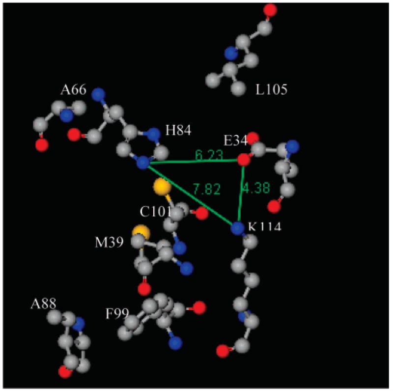
Figure 11.

Arrangement of amino acid residues, considered for ANS binding, in the crystal structure of TL (PDB 1XKI (18)). Residue Leu105 is modeled from the solution structure of TL by SDTF (20). Gray, blue, red, and yellow balls represent carbon, nitrogen, oxygen, and sulfur atoms, respectively. The numbers represent distances between the atoms (Å). The image was generated by ViewerLite 5.0 (Accelrys Inc.).
