
Fig. 1. In vivo differentiation potential of murine iPS cells.
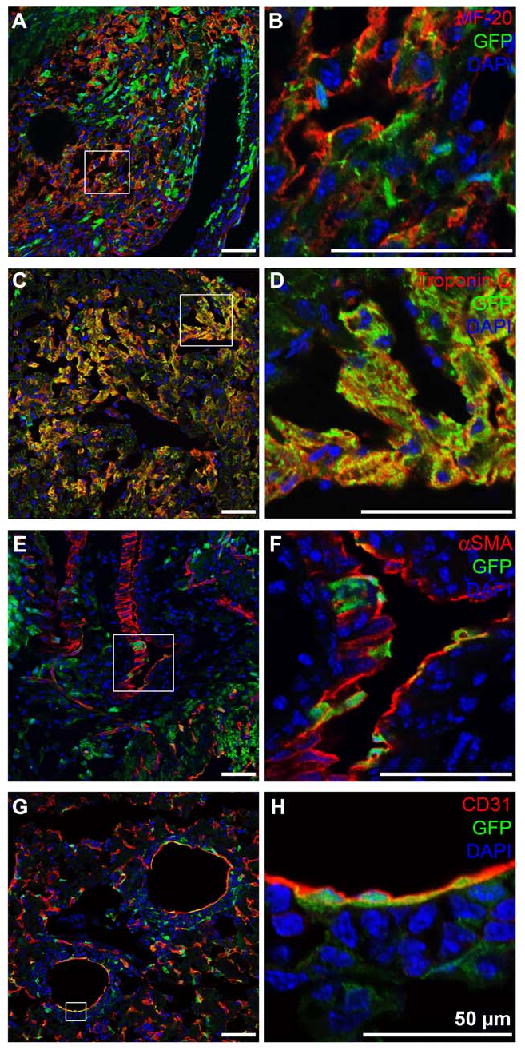
Immunohistochemical double staining of heart sections of newborn 2D4 iPS cell-derived chimera mice shows that GFP-expressing iPS cells (green) contribute to cells of the cardiovascular lineage including cardiomyocytes: (A, B) MF-20 (red), (C, D) Troponin C (red); smooth muscle cells: (E, F) αSMA (red); and endothelial cells: (G, H) CD31 (red). DAPI-staining was performed to show cell nuclei (blue). Scale bars equal 50 μm.