
Figure 4.
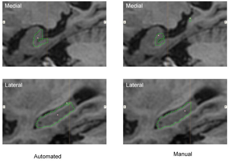
The largest outlier of the automated hippocampal segmentation in the subset of 15 subjects in terms of difference compared with manual measures. This difference in volume can be largely attributed to the automated region including more of the tail of the hippocampus (lower panel) and more of the medial aspect of body and tail (upper panels) than was included in the manual segmentations.