
Figure 5.
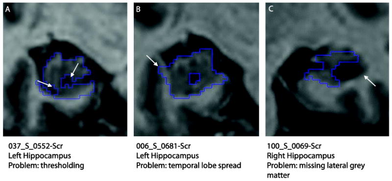
Automated hippocampal segmentation errors. (A) Thresholding excluding hippocampal tissue, (B) extra-hippocampal tissue included (white and grey matter of the temporal lobe) and (C) exclusion of lateral hippocampal grey matter due to large hippocampal cyst and lack of temporal horn.