Figure 6.
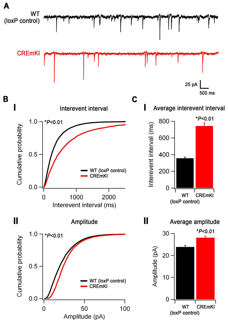
Neuronal activity-dependent Bdnf expression controls the development of cortical inhibition. A) Representative traces of mIPSCs recorded from layer II/III V1 pyramidal neurons in loxP control and CREmKI acute cortical slices. B) Cumulative probability distributions of mIPSC interevent intervals (I) and amplitudes (II) recorded from loxP control and CREmKI neurons. P<0.01 by either Kolmogorov-Smirnov test or Monte Carlo simulation (see Supplemental Methods). C) Average interevent interval (I) and amplitude (II) of mIPSCs recorded from loxP control and CREmKI neurons. Data are mean ± SEM, P<0.01 by Student’s t-test. Data are from 18–20 cells/genotype recorded from 12 pairs of littermates.

