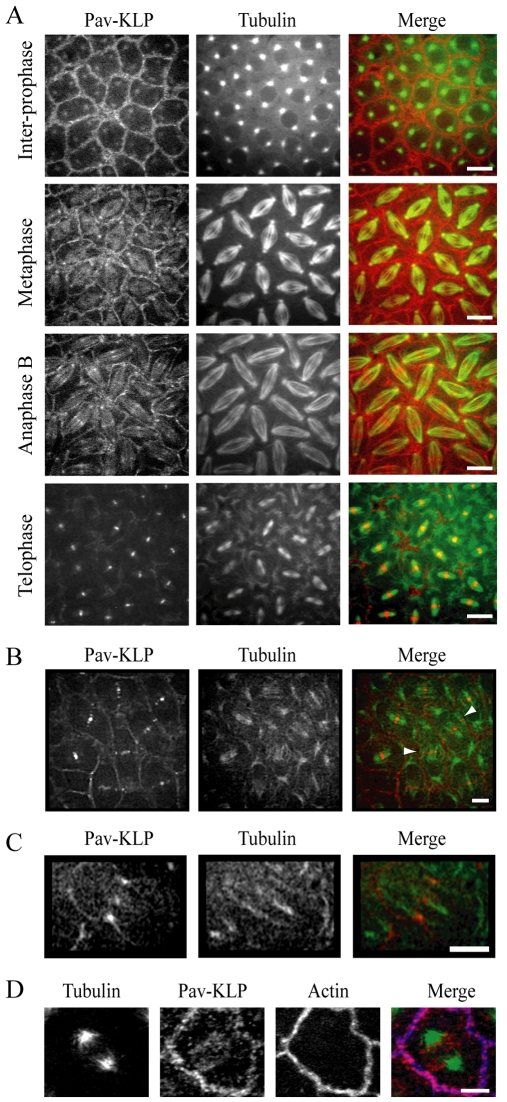
Fig. 1.
Pav-KLP localization. (A) In the syncytial embryo, GFP–Pav-KLP localizes to the centrosomes and the cortex before NEB. After NEB, GFP–Pav-KLP also localizes to spindle microtubules. It concentrates at the equator during anaphase B and the midbody during telophase. In the merged image, GFP–Pav-KLP is red and tubulin green. Scale bars: 10 μm. (B) GFP–Pav-KLP distribution during early telophase. Pav-KLP (red) is mainly focused in the narrow band of MTs (green) engaged in forming the spindle midbody (‘internal’ MTs), although a small fraction is on MTs positioned outside the spindle midbody and contacting the cortex (‘peripheral’ MTs, arrowheads). Scale bar: 10 μm. (C) Initial stages of furrow ingression in GFP–Pav-KLP post-cellularized embryos. Pav-KLP (red) accumulates on the spindle (green) at the site of furrow ingression. Scale bar: 10 μm. (D) Immunolocalization of Pav-KLP in the syncytium. Pav-KLP (red) at the cortex colocalizes with actin (blue). Regions of colocalization are pink. Tubulin is shown in green. Scale bar: 2 μm.

