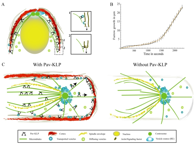
Fig. 6.
Model for Pav-KLP function. (A) Qualitative model of furrow growth during cellularization: membrane vesicles originate near the centrosomes, diffuse through the cytoplasm, are transported by Pav-KLP on astral MTs and, upon reaching the furrow, are instantly fused laterally with it (inset 1). Pav-KLP bound to the furrow at the tip moves towards the MT plus end, pulling the furrow down (inset 2). (B) Model prediction for the furrow length as a function of time (two black curves). At early times the curve is derived from solving the membrane-unfolding equations; at later times the curve is derived from solving the vesicle-transport equations. These curves fit the experimental data (orange). Bars show experimental errors. (C) Pav-KLP functions on a single spindle in the syncytium. The cartoon shows a top view of a single spindle surrounded by the mitotic furrow. Inside the spindle envelope (yellow), Pav-KLP crosslinks ipMTs and helps stabilize the overlapping MTs in the central-spindle region; outside the spindle envelope, Pav-KLP localizes on the astral MTs, moves towards the cortex and promotes furrow growth (perpendicular to the plane shown) by transporting actin, vesicles or signaling factors to the cortex. In the absence of Pav-KLP (right), ipMTs inside the spindle envelope are not bundled and the spindle loses its shape; whereas outside the spindle envelope, loss of Pav-KLP causes defects in actin distribution, inhibiting the growth of the mitotic furrows.

