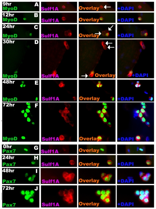
Fig. 2.
Sulf1A activation in satellite cells precedes asynchronous MyoD activation in vitro. Satellite cells on dissociated single fibres stained after different time intervals in vitro for MyoD (column 1, A-F) or Pax7 (column 1 G-J) and Sulf1A (column 2) using a double immunofluorescence procedure. The superimposed images (overlay) of MyoD and Sulf1A as well as Pax7 and Sulf1A staining are shown in column 3 with the addition of DAPI staining in column 4. Some satellite cells, indicated by arrows at the 9-, 24- and 30-hour time points show Sulf1A staining but still no MyoD staining. hr, hours.

