
Figure 1.
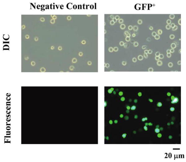
Fluorescence of HL-60 cells transfected with lentivirus containing green fluorescent protein (GFP) or not (negative control). A, Differential interference contrast (DIC) and fluorescence microscopy of individual, transfected cells. The same field of view is shown by DIC above and fluorescence below. Total original magnification, ×400. GFP+, GFP positive.