
Figure 3.
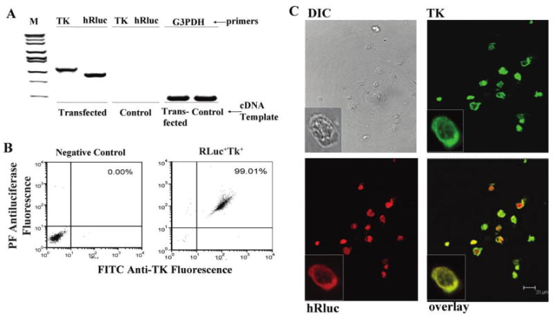
High level of expression of both the luciferase bioluminescence marker and herpes simplex virus (HSV) thymidine kinase (TK) suicide trap in dually transfected cells. A, Expression by reverse-transcriptase polymerase chain reaction (RT-PCR) of luciferase and HSV TK in dually transfected HL-60 cells or control HL-60 cells. B, Flow cytometry of dually transfected or control HL-60 cells stained intracellularly for both luciferase and TK protein expression. Flow was conducted after 2 months of growth in no selection media. C, Confocal microscopy of dually transfected or control cells stained for expression of luciferase and TK, or visualized by differential interference contrast optics. cDNA, complementary DNA; DIC, differential interference contrast; G3PDH, RT-PCR using primers for this as a constitutive control; hRluc, RT-PCR using primers specific for luciferase; M, molecular weight markers; PE, phycoerythrin; TK, RT-PCR using primers specific for HSV TK.