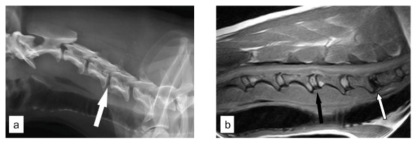
Figure 3.
a. The radiographic examination of the cervical spine in a Dachshund with a surgically confirmed disc extrusion at C4-C5 (white arrow). Notice the absence of CDVR at C4-C5.
b. A T1-weighted sagittal magnetic resonance image of the cervical spinal cord of the Dachshund in 3a. Notice the extent of calcification of the C6-C7 disc (white arrow) compared to the affected disc at C4-C5 (black arrow).

