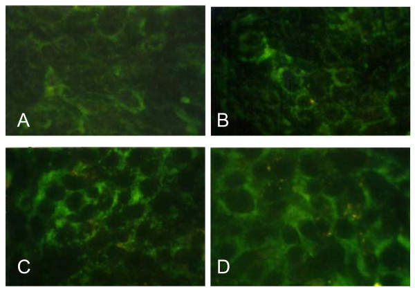
Figure 4.
Immunofluorescence staining of Bax protein in MCF-7 cells treated with 9 μg/ml 3HFD. (A) Untreated MCF-7 cells at 0 h of 3HFD treatment showed a low level of Bax protein. (B), (C) and (D) MCF-7 cells after 6, 12 or 24 h of 3HFD treatment exhibited a marked increase of immunofluorescence for Bax protein.

