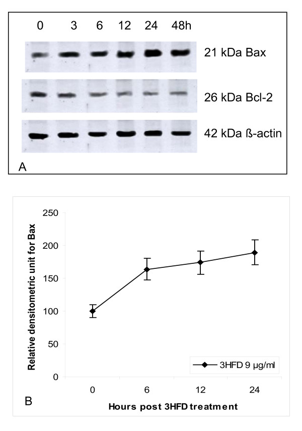
Figure 5.
Western blot analysis of Bax protein in MCF-7 cells treated with 9 μg/ml 3HFD. (A) MCF-7 cells treated with 9 μg/ml 3HFD for the indicated times were resolved on a 15% PAGE and subjected to Western blotting. Bax protein expression increased as early as 3 h post treatment, whereas Bcl-2 levels were not altered and remained low throughout the experiment. (B)The intensity of Bax protein expression increased in a time-dependent manner. Results are presented as the mean ± SEM of 3 independent experiments.

