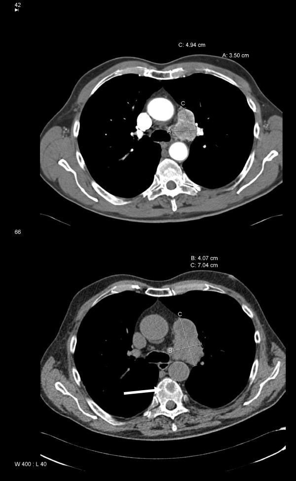
Figure 2.
Computed tomography of the chest showing mediastinal lymph node enlargement (upper image: September 2005, i.e. initial diagnosis of metastases). Slow progression in the absence of treatment (lower image: June 2008, i.e. before initiation of sunitinib therapy). The white arrow indicates metastasis in a thoracic vertebra.

