Figure 1.
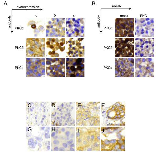
Immunohistochemical stainings of primary breast cancer and validation of antibody specificity. MCF-7 cells were transfected with vectors encoding PKCα (α), PKCδ (δ), or PKCε (ε) (A) and MDA-MB-231 cells were mock-treated or transfected with siRNA targeting PKCα (α), PKCδ (δ), or PKCε (ε) (B). Pellets of transfected cells were arranged in a cell line array and immunohistochemistry was performed with antibodies towards indicated PKC isoforms. (C-J) Examples of immunohistochemical staining of PKCα in breast cancer specimens from cohort II showing negative (C and G), low (D and H), moderate (E and I), and intense (F and J) staining with 20× magnification (C-F) and with 40× magnification (G-J).

