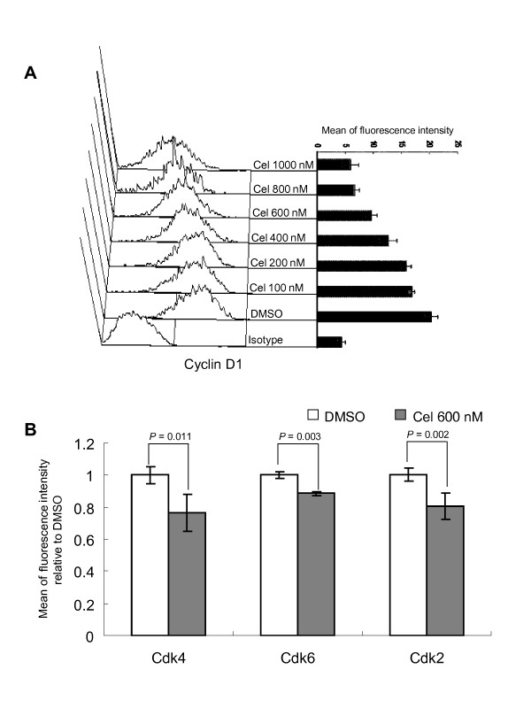
Figure 3.
Celastrol decreases the level of Cyclin D1 and some Cdks. Following treatment, cells were incubated with indicated antibodies and the expressions of proteins were detected by FCM detailed in Methods. A: Celastrol induces reduction of Cyclin D1 in a dose-dependent manner. The left panel shows the histogram for FCM detection of Cyclin D1 expression with X-axis as fluorescence intensity and Y-axis representing cell number. The right panel shows the detected intensities of this protein. Each value represents the mean of three independent experiments. B: Effects of celastrol on Cdk4, Cdk6, and Cdk2 expressions. After exposure to 600 nM celastrol for 1 d, the proteins were detected by FCM. Y-axis represents the relative levels of each protein in different treatments, with the protein level in DMSO-control sample being set at 1.0. Each value is the mean of three independent experiments.

