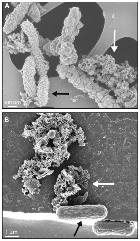
Fig. 1.
Comparison of scanning electron micrographs of circum-neutral pH Fe(II)-oxidizing bacteria. A) Nitrate-reducing Fe(II)-oxidizing Acidovorax sp. strain BoFeN1 cells heavily encrusted in iron(III) (hydr)oxides (black arrow) plus extracellular Fe(III) mineral precipitates (filaments – white arrow). B) Phototrophic Fe(II)-oxidizer Thiodictyon sp. strain F4 (black arrow) associated with, but not encrusted in iron(III) (hydr)oxides (white arrow). Thiodictyon sp. strain F4 was used in the Fe-isotope study of phototrophic Fe(II)-oxidizers by Croal et al. (2004).

