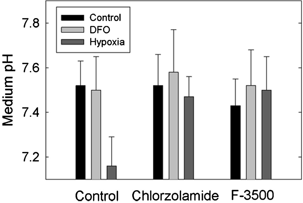
Figure 8.
Effect of CA Inhibitors on medium pH. Cells were treated 2 days post plating with DFO, or exposed to 1% oxygen, for 16 hr in the absence or presence of 100-µM chlorozolamide or F3500. The pH of the medium was tested at the end of the incubation, which will be noted is higher, overall, than that in Figure 7 because of the lower density of the cells. Data are reported as the mean ± SD, n = 3.

