
Figure 2.
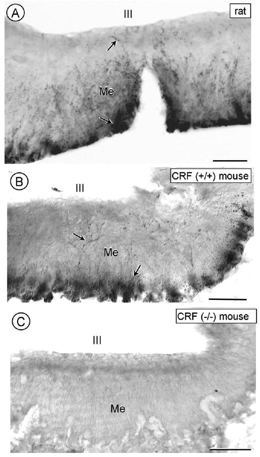
Light microscopic images showing immunoperoxidase labeling for CRF in the hypothalamic median eminence of a rat (A), a CRF (+/+) mouse (B), and a CRF (−/−) mouse (C). The CRF-immunoreactivity is seen in varicose processes (arrows) mainly in the outer but also inner layer of the median eminence (Me) below the third ventricle (III). No similar labeling is seen in a CRF (−/−) mouse. Scale bars = 80 mM.