
Figure 4.
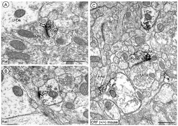
Electron micrographs showing the predominant dendritic immunoperoxidase labeling for CRFr in the CeL. The peroxidase reaction product is seen in the region of the postsynaptic membrane specialization in a dendritic spine (r-Sp) and small dendrite (r-De) in A and B, respectively. Unlabeled dendrites (U-De) are seen in the neuropil. In C, aggregates of immunoreactivity are seen on and near a restricted portion of the plasma membrane in a CRFr-labeled dendrite (r-De) and in a dendritic spine (r-Sp). Immunolabeling is also seen in small axons (I-Ax) and a glial process (*). Block arrow, plasmalemmal labeling in dendritic and glial profiles; U-Te, unlabeled terminal; U-Sp, unlabeled spine; Ax, unlabeled axon. Scale bars = 0.5 μm.