
Figure 8.
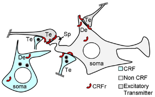
Schematic diagram showing the predominant, but not exclusive, postsynaptic distribution of CRFr (red arc) at asymmetric excitatory-type synapses on the spine (sp) and dendrite (De) of a non-CRF-containing neuron in the CeA. The asymmetric synapses are typical of afferent inputs from terminals that contain excitatory transmitters. The CRF labeling is depicted in a separate neuron, whose soma, dendrite (De), and terminals (Te) show a more sparing distribution of the CRFr. The black circular symbols in the CRF De and Te are peptide-storage vesicles.