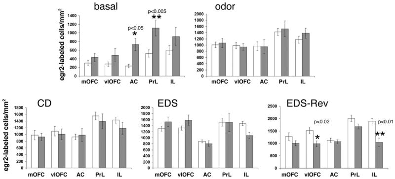
Fig. 5.
Comparison of the densities of egr-2-labeled cells in the OFC and mPFC of wild type and D2 mutants at baseline, after odor exposure alone, and after completion of the CD, EDS, or EDS-Rev phases of the ASST. White bars: wild type. Gray bars: D2 mutants. The individual subregions of the OFC (mOFC, vlOFC) and the mPFC (AC, PrL, IL (layers II/III)) are indicated. Data are means ± SEM of determinations made from five to seven animals per group and genotype. egr-2 Densities for D2 mutants at baseline, after odor exposure, and after CD- and EDS-Rev-testing are those also shown in Fig. 4. The P-values indicated reflect the differences between measures of D2 mutants and wild type in the same anatomic region under the same test condition (two-tailed t-tests).

