Fig. 1.

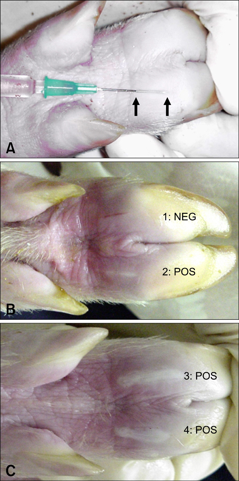
(A) Intradermal inoculation in the heel bulb. The space between the two arrows marks the portion of the needle that lays within the dermis, approximately 1.2 cm. Inoculums were released while slowly removing the needle. (B and C) Replication of O/UKG/35/01 at the inoculation site 24 h after intradermal inoculation with 700 PFU/5 µL (1 and 2) and 70,000 PFU/5 µL (3 and 4). The presence of a vesicle (POS) indicates a positive result. The absence of a vesicle (NEG) indicates a negative result.
