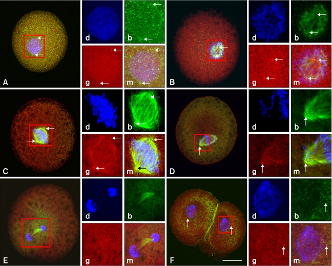
Fig. 3.
Nuclear progression and microtubule organization in the first mitotic phase of bovine SCNT embryos. Nuclear progression and microtubule distribution in the different phases of the first mitosis of SCNT embryos were examined 20 h after fusion. (A) Interphase. Two γ-tubulin foci are seen around a pronucleus-like structure. (B) Prophase. (C) Prometaphase. (D) Metaphase. (E) Ana-telophase. γ-tubulin foci were not detected. (F) Two-cell stage, One γ-tubulin focus is seen around the nucleus of both blastomeres. Insets indicate DNA (d; blue), β-tubulin (b; green), γ-tubulin (g; red; arrows), and merged (m) images. Scale bar = 50 µm.

