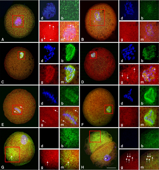
Fig. 4.
Abnormal nuclear and microtubule configuration in the first mitotic phase of bovine SCNT embryos. Microtubule distribution in the SCNT embryos was examined 20 h after fusion. (A) Multiple and multipolar γ-tubulin foci were organized near the nucleus of SCNT embryos. (B) Chromosomes were condensed but mitotic spindles were not organized. (C) Mitotic spindles were organized without any spindle poles. (D) Mitotic spindles were organized from multiple poles. (E) Two γ-tubulin foci were seen near the mitotic spindle but did not connect with them. (F) Three mitotic spindles were organized without any spindle poles. (G) A cytoplasmic microtubule network from a focus (within square) was organized at the opposite pole of the nucleus. (H) Cytoplasmic fragment without a nucleus. Insets indicate DNA (d; blue), β-tubulin (b; green), γ-tubulin (g; red), and merged (m) images. Scale bar = 50 µm.

