
Figure 3.
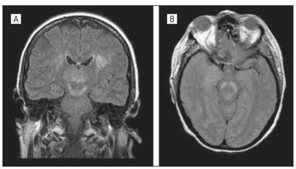
Axial proton density magnetic resonance image (left) and coronal fluid-attenuated inversion recovery magnetic resonance image of patients with West Nile virus encephalitis showing increased signal in the upper brainstem, thalamus, and basal ganglia (A) and substantia nigra of the midbrain (B).