
Figure 1.
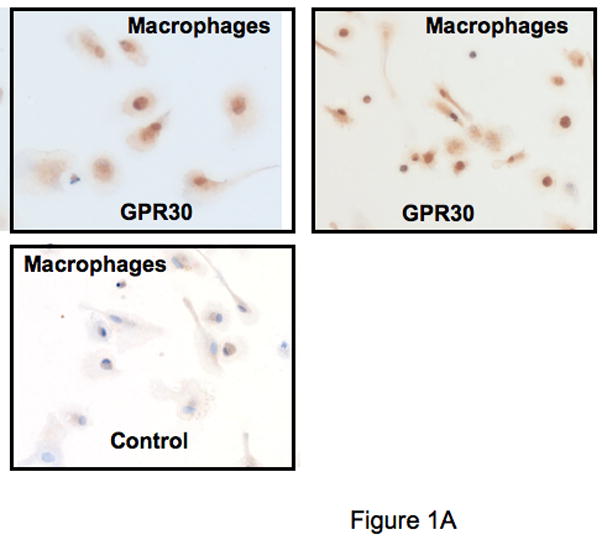
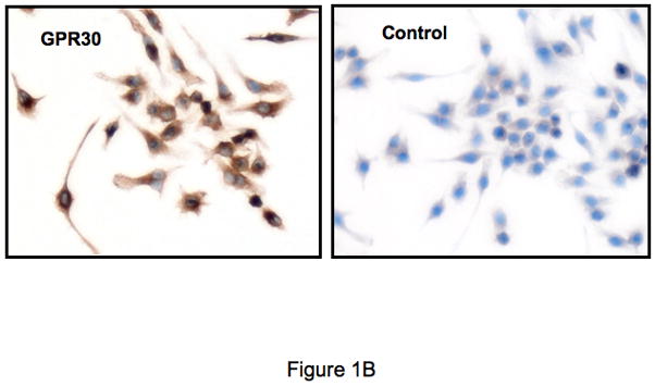
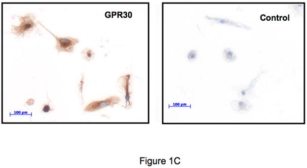
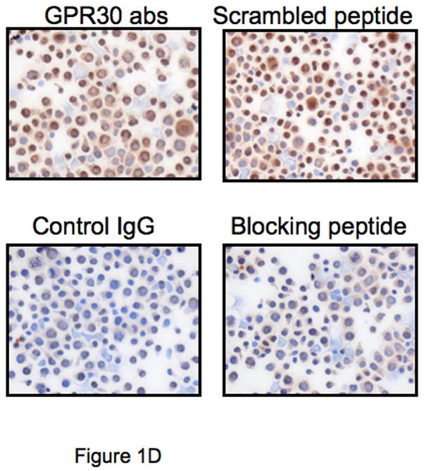
GPR30 expression by immunohistochemical staining (brown) in primary immune cells and cell lines. (A) Human macrophages, (B) Mouse RAW 264.7 cells, (C) Rat microglia and (D) Human regulatory T cells all stain with an antibody against GPR30. Staining is primarily cytoplasmic in the mouse and rat cells, while in the human macrophages and T cells it is both cytoplasmic and nuclear. There is little to no staining with a control antibody. In addition, staining is abolished by preincubation with a C-terminal peptide to GPR30 but not with a scrambled version of the same peptide (D). Nuclei are lightly counterstained with hematoxylin. All images taken with a 63- oil objective on a Zeiss Axioskop. Scale bars = 100 μm in D.