
Figure 5.
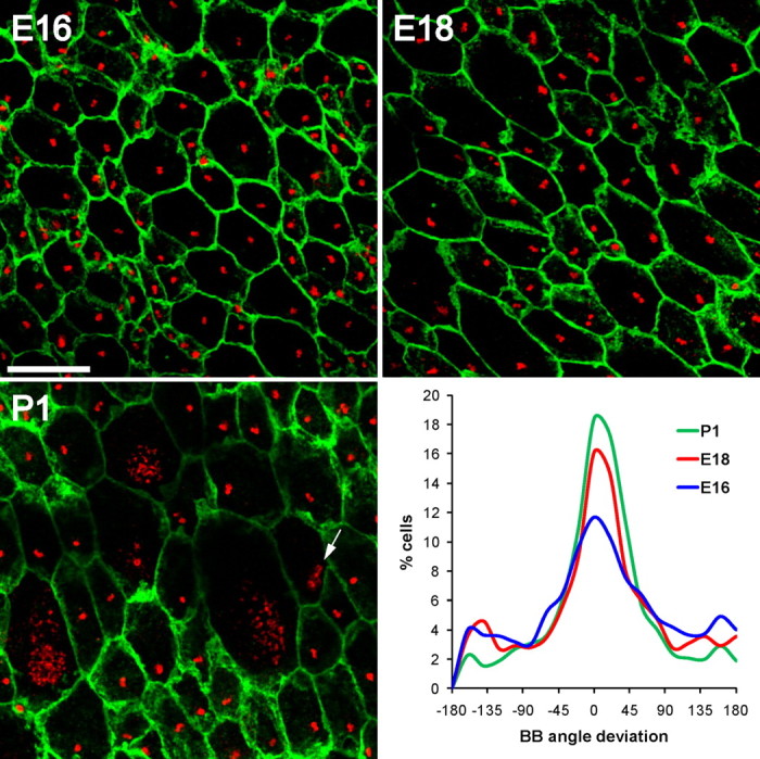
Planar polarity of basal body position in radial glia. Confocal images of lateral wall wholemounts from E16, E18, and P1 wild-type mice stained for β-catenin (green) and γ-tubulin (red). Images were taken from a mid-ventral region of the wholemount near the foramen of Monro (supplemental Fig. S9, available at www.jneurosci.org as supplemental material, arrowhead). At successively older ages, radial glial apical surfaces expanded and by P1, some radial glia had already transformed into ependymal cells with multiple basal bodies. Note that some cells had dense punctate γ-tubulin staining likely corresponding to deuterosomes (arrow in P1 image). The histograms show that at successively older ages, an increasingly larger fraction of radial glia exhibited planar polarity of their single basal body. Neighboring cells had their basal body displaced to the ensuing “downstream” side of the cell with respect to CSF flow. Scale bar, 10 μm.