
Fig. 5. GSK-3β inhibitors attenuate radiation-induced apoptosis in IEC-6 cells.
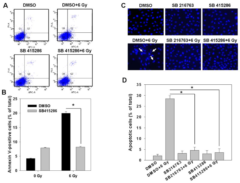
IEC6 cells were treated with DMSO, 10 μM SB216763 or 25 μM SB415286 16 h prior to irradiation with 6 Gy. At 24 h after irradiation, cells were stained with Annexin V-APC/propidium iodide and analyzed by flow cytometry (A, B). Alternatively, cells were fixed and stained with DAPI, and apoptotic cells indicated by arrows were counted in multiple randomly selected fields (C, D). Shown are representative diagrams of distribution of stained cells (A), representative micrographs (C) and bar graphs of the average percent of apoptotic cells vs. total cell number for each treatment with SEM from three experiments; *, P<0.05 (B, D).