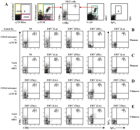
Figure 8. NKT cells differentially express the CD8α and CD8β chain.
(A) The experimental and analysis scheme for detecting total and co-receptor CD8α- and CD8β-expressing NKT cells. (B) and (C) NKT cells in PBMCs from healthy latent EBV-infected subjects [EBV+(La)], IM patients at year 1 post-onset [EBV+(IMy)], EBV-associated HL patients [EBV+(HL)] and EBV-negative normal control subjects (NS) were assessed by flow cytometry using the gate of either CD1d tetramers vs. anti-αβTCR mAb (B) or anti-Vα24 mAb vs. 6B11 mAb (C). Further dot plot analysis of CD8α vs. CD8β in gated NKT cells was shown. (D) and (E), NKT cells in thymus (Thy) and liver (Liv) from hu-thy/liv-SCID chimeras challenged i.t. with EBV (EBV+) or unchallenged (EBV−) were assessed by flow cytometry using the gate of either CD1d tetramers vs. anti-αβTCR mAb (D) or anti-Vα24 mAb vs. 6B11 mAb (E). Further dot plot analysis of CD8α vs. CD8β in gated NKT cells was shown. Data were representatives of 5 similar experiments in each group.

