Figure 4.
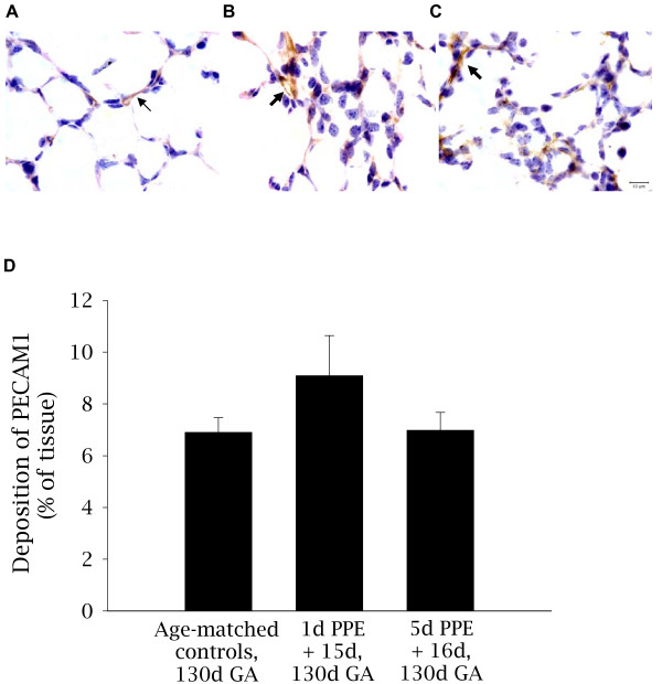
Localization and relative abundance of PECAM1 in control and embolized fetal lung tissue. Light micrographs depicting the localization of PECAM1 (brown staining); a marker of endothelial cells, in control (A), 1d PPE + 15d (B), and 5d PPE +16d (C) lung tissue. Nuclei are counterstained blue with haematoxylin. The thin arrow indicates normal pulmonary capillaries (A) and the thick arrows indicate abnormal capillary staining in the parenchyma of 1d PPE + 15d and 5d PPE +16d fetuses (B-C). The scale bar in (C) represents 10 μm in all three images. The relative abundance of PECAM1 (D) was not significantly different between groups. However, the PPE fetuses appeared to have larger capillaries compared to control fetuses and PECAM1 staining was less common within the septal walls of PPE fetuses.

