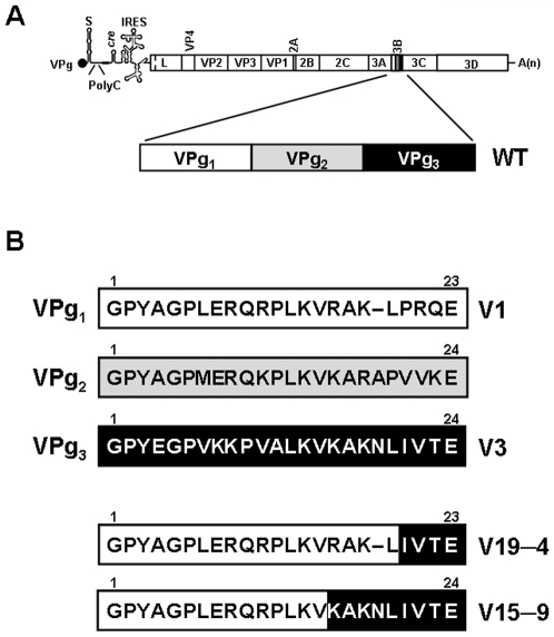
Figure 1. Schematic representation of the FMDV genome and of the constructions with one copy of VPg.
A, FMDV genome (VPg is the protein covalently linked to the 5′–end of the RNA; PolyC is the internal polycytidylate tract; IRES is the internal ribosome entry site; A(n) is the PolyA at the 3′–end). The genomic region encoding the viral polyprotein is boxed. The viral polyprotein is processed into the different mature proteins indicated in each corresponding box (based in [15]). Gene 3B (highlighted) encodes 3 different but related copies of FMDV protein–primer VPg. B, Amino acid sequence of VPg1, VPg2 and VPg3 of FMDV C–S8c1. Infectious FMDV clones were constructed either to express VPg1 (V1), VPg3 (V3), a chimeric VPg consisting of the first 19 residues from VPg1 (N-terminus) and the last 4 residues from VPg3 (C-terminus) (V19–4), or a chimeric VPg containing the first 15 residues from VPg1 and the last 9 residues from VPg3 (V15–9). The starting pMT28 plasmid and procedures for the construction are detailed in Materials and Methods.

